The Russian sturgeon, Acipenser gueldenstaedtii Brandt & Ratzeburg, 1833, is an ancient fish native to Black Sea, Sea of Azov and Caspian Sea entering all the main rivers that empty into them.This species has both anadromous and freshwater populations: at sea, it occurs in shallow coastal and estuarine zones, while in freshwaters it inhabits the deep parts of large rivers with moderate to swift current. It was introduced throughout Europe and, due to loss of habitat caused by the construction of dams and to overfishing to collect its eggs, it is now considered critically endangered although it is a promising species for aquacultural purposes.
In recent decades, sturgeon farming for meat and caviar production has increased in Europe. Nevertheless, the development of sturgeon aquaculture activities has been frequently accompanied by disease outbreaks, including those unknown prior to farming. Unfortunately, research on the major sturgeons diseases had not received much attention in the past and, therefore, the knowledge on their prevalence, distribution, origin and ecological significance is still fairly limited. Although sturgeons are considered relatively resilient to diseases, several bacterial, viral and parasitic diseases have been reported worldwide, including Columnaris disease, motile Aeromonas septicemia, yersiniosis, pseudomonosis, epitheliocystis, white sturgeon adenovirus, herpesvirus and iridovirus.
To date, no reports of skin and/or oral tumor-like masses in sturgeons associated with mycobacterial infections are documented. Only few papers described a group of Siberian sturgeons (Acipenser baeri) imported in Italy from France showing visceral granulomas, and caviar fishes from Iran exhibiting gill lesions, both caused by Mycobacterium marinum.
The non-tuberculous mycobacteria (NTM), also known as environmental mycobacteria (EM), atypical mycobacteria (AM) and mycobacteria other than tuberculosis (MOTT), include those Mycobacterium species which are not members of the Mycobacterium tuberculosis complex (and M. leprae). NTM have a worldwide distribution as saprophytes in soil and in treated/untreated waters, where they can remain viable for over 2 years. NTM include many species of acid-fast bacteria, some of which are capable of causing chronic progressive diseases in mammals, birds, reptiles and fish. Thus, infected fish may be considered as primary reservoir for these pathogens and infection is probably acquired from NTM present in the environment by ingesting contaminated food or water containing organic matter, or through gill or skin lesions caused by trauma and parasites. Mycobacteria may cause cutaneous lesions or spread into internal organs through the circulatory or lymphatic systems. Transovarian transmission has also been documented in Siamese fighting fish (Betta splendens). All fish species should be considered susceptible to mycobacteriosis, with a higher prevalence in farmed animals and significant losses frequently observed in aquacultural activities. However, mycobacteriosis infections and mass mortalities in wild fish is not easy to recognized but see, because infected fish die and are easily predated or scavenged.
Disease outbreaks in farmed fish can be related to several management features, such as the quality and quantity of food and water supplied, and the rearing density. As a result, inadequate management could cause abnormal stress associated with a decrease of the normal resistance of the host.
Mycobacteriosis in fish is currently attributed to a wide array of species belonging to the Mycobacterium genus, but historically M. chelonae, M. fortuitum, and M. marinum are the most reported species. Furthermore, M. marinum is the most common in a wide range of saltwater fish and M. chelonae (M. salmoniphilum) has been identified in different coldwater fish, especially in salmonids.
In general, piscine mycobacteriosis is a systemic subacute to chronic progressive disease causing grey-whitish nodules (granulomas) and enlargement of various organs (mainly spleen, liver and kidney). Clinical signs are often lacking and hardly detectable until advanced stages, in which cachexia, lordosis, pale gills and skin ulcerative lesions can be seen in some fish species. Skin granuloma caused by NTM infection was not reported in any fish species, except for Atlantic salmon farmed in British Columbia.
However, to our knowledge, no skin or oral granulomatous inflammation with excessive mineralization associated with mycobacteria has ever been observed in sturgeons. The aim of this study, therefore, was to describe in detail the first occurrence of tumor-like lesions associated with Mycobacterium chelonae in the Russian sturgeon, Acipenser gueldenstaedtii.
Results
Macroscopical findings
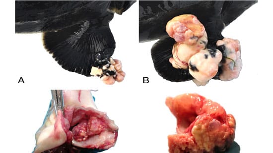
At postmortem examination, a total of 20 skin and oral cavity masses were found in the 15 sturgeons examined. The 73 per cent of the fish exhibited single lesions, mostly located in the mouth (82 per cent), although 27 per cent of the sturgeons showed multiple skin and oral masses. Total skin lesions accounted for 35 per cent, and were located in both pectoral (43 per cent) and caudal fins (57 per cent). These lesions were mostly medium-sized (Figure 1A), except for one large (Figure 1B) found in case No. 7 and another small one (case No. 1) located in the pectoral and caudal fins, respectively (Table 1). Grossly, all the skin masses were firm, whitish in color and showed a cauliflower-like exophytic growth (Figure 1A and B). Oral cavity masses (Figure 1C) accounted for 65 per cent and were significantly larger (>2 cm) compared to those on the skin (Kruskal-Wallis H = 12.504, P < 0.01; Dunn’s post-hoc test, mouth vs caudal fin Q= 2.622, P < 0.05). Oral masses were mostly infiltrative within the submucosa and, on cut section, were composed of a yellowish gritty material (Figure 1D). No one of the sturgeons examined showed visceral lesions.
Cytopathology and histopathology
Grossly, unstained smears showed a chalky, white material. Cytologically, the aspirate exhibited a large amount of amorphous granular, dark-gray to bluish material admixed with glassy retractile fragments, suggestive of calcium salt deposit, uniformly dispersed in the background. Cellular components were represented by a moderate to severe mixed inflammatory reaction (macrophages, lymphocytes and multinucleated giant cells) and numerous reactive spindloid, fibroblast-like cells. There was no evidence of bacteria or other infectious agents and ZN was negative for acid-fast bacilli.
On histopathology, skin masses showed generally a multinodular aspect, with an infiltrative growth pattern into the dermis and subcutis, surrounded by abundant fibrous stroma. Only one caudal fin skin mass (case No. 1) exhibited a nodular pattern and was well encapsulated and demarcated (Figure 2A).
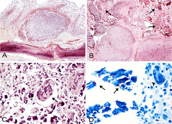
Skin lesions were mostly characterized by mild to moderate deposition of amorphous granular calcium material, positive to Von Kossa stain, and by mild to moderate inflammations composed mostly of macrophages, lymphocytes, and few giant cells. Mild to moderate fibroplasia was also evident with Masson’s Trichrome stain. Oral masses were characterized by a multinodular infiltrative pattern into the submucosa and were poorly demarcated and nonencapsulated (Figure 2B). Moderate to severe inflammatory reaction with a high number of macrophages, lymphocytes and numerous giant cells, phagocytizing calcium debris were found (Figure 2C). Excessive mineralization deposit associated with severe fibroplasia and necrosis was also observed. Red, rod-shaped extracellular acid-fast bacteria within mineralized deposits of calcium and necrotic debris were seen in one of the oral masses (case No. 3) examined (Figure 2D).
Fourteen out of twenty lesions (70 per cent), mostly found in oral cavity (86 per cent), exhibited severe histological changes, and showed a higher degree of severity than those located in the skin (Kruskal-Wallis H = 11.020, P < 0.01; Dunn’s post-hoc test, mouth vs caudal fin Q= 2.578, P < 0.05). Spearman correlation analysis found a significant positive correlation only between the size of the lesions and their histopathological severity (ρ = 0.7028, P < 0.001).
Bacteriological and molecular results
None of the smears obtained from the masses sampled were positive to ZN stain. Colonies growth from cultured oral and skin masses, were clearly visible in Löwenstein- Jensen media tubes showing acid-fast bacilli at ZN stain (both tubes incubated at 25°C and 37°C were positive). These colonies were phenotypically and biochemically identified as Mycobacterium chelonae. All the internal organs cultured were negative for mycobacteria. Furthermore, no colony growth of mycobacteria were observed from all negative controls.
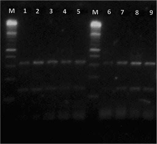
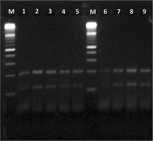
All the strains subjected to PCR-RFLP analysis showed a restriction pattern identical to the one of M. chelonae (Figures 3 and 4), confirming the results obtained by phenotypic and biochemical identification.
Discussion
In the present study, the occurrence of Mycobacterium chelonae associated with tumor-like masses with excessive calcium deposition was described for the first time in farmed sturgeons. In general, the presence of this mycobacterium species and its association with gross lesions is considered quite rare in fish, with few reports in the literature. These records described M. chelonae mostly associated with granuloma-like visceral nodules in farmed salmon and turbot, in wild perch, and also in ornamental fish. Furthermore, M. chelonae was evidenced in several other wild and domestic animals but mostly associated with ulcerative skin lesions and references therein.
In all the sturgeons examined in this study, the lesions were different in size and larger masses were mostly observed in the oral cavity, most of which showing a severe histological pattern significantly correlated with their size and with an infiltrative activity. Moreover, all the skin and mouth lesions were characterized by different severity levels of chronic granulomatous inflammation. To the best of our knowledge, granulomatous infiltrates associated with few bacteria, isolated and confirmed by PCR as M. chelonae, were described in salmon. It is worth noting that none of the lesions we observed showed typical granulomas, although these structures are quite common in fish organs with mycobacteriosis and references therein. In particular, typical granulomas are characterized by peripheral fibrosis and several layers of epithelioid cells surrounding necrotic areas containing a large amount of acid-fast bacilli. Conversely, we observed scant ZN positive bacilli intermingled with abundant mineralization, tissue necrosis and fibroplasia. Strong positivity to Von Kossa stain of mineralized material confirmed that calcium salt deposition was diffusely present within nodular and multinodular masses and increased from smaller to larger lesions. In general, mineralization is rarely encountered in piscine mycobacteriosis and references therein. Nevertheless, ectopic mineralization in soft tissues has been reported for many other fish species and references therein, although different names to describe similar lesions have been used, leading to confusion in terminology. In particular, an ectopic mineralization characterized by tumorlike nodules in subcutaneous tissues containing calcium deposition is defined as Calcinosis circumscripta (CC) and references therein.
Thus, due to the macro and microscopic aspect of the masses we examined (that were all characterized by single or multiple nodules containing abundant calcium deposits), their distribution (single or multiple) and localization (skin and oral mucosa), CC syndrome was considered as a possible diagnosis and references therein. This syndrome is a disorder affecting humans and several animal species (but never described in sturgeons before) and is classified in 3 different forms: metastatic, idiopathic and dystrophic calcinosis and references therein.
Aetiopathogeneses for calcinosis include trauma, foreign body reaction, neoplasms, connective tissue diseases, abscesses, granulomas, inherited disorders and infections. Generally, the ectopic calcified masses consist of hydroxyapatite and amorphous calcium phosphate. It has been hypothesized that the denatured proteins of necrotic cells can serve as a core for ectopic calcification, and that the following degeneration of the collagen fibers and subcutaneous fat tissue can induce the calcification process. In addition, inflammation and fibrosis may be frequently observed as a reaction of calcified deposits (e.g. foreign body reaction).
From a clinical point of view, nodules of calcium deposits can cause skin ulcerations occurring in areas of recurrent microtraumas. Thus, most probably, the calcinosis we observed in the sturgeon masses was of dystrophic origin, because tissue injures especially occurred over body prominences (e.g. pectoral and caudal fins), or in areas susceptible to trauma (e.g. mouth). Probably, therefore, sturgeons affected by these masses can be assumed to have feeding problems, leading to starvation.
Furthermore, the fact that a number of sturgeons exhibited mouth and skin lesions could be related to farming conditions and, in particular, to stressful breeding density. As a consequence, the mycobacteria observed inside the sturgeon masses could have been present in the rearing environment (i.e. the water) and only after traumatic events infected the fish lesions. Based on this hypothesis, we can suppose that the granulomatous inflammation process could be related to the presence of a foreign body reaction (i.e. the calcium), and the occurrence of M. chelonae associated with tumor-like skin and oral lesions was only a secondary event. In fact, this mycobacterium is a fast growing, opportunistic bacillus ubiquitous in nature, exceptionally pathogenic and mainly associated to chronic granulomatous infections in immunocompetent and immunosuppressed subjects. The histological pattern observed in almost all the sturgeon lesions (19 out of 20; 95 per cent) seems to confirm this hypothesis, because no acid-fast bacilli were noticed in the macrophages cytoplasm, in epithelioid cells, nor in multinucleated giant cells which contained almost only calcium. Only in one of the oral lesion examined, we found a few extracellular acid-fast bacteria intermixed calcium deposits. However, a number of studies demonstrated that the sensitivity of some piscine mycobacteria to ZN staining could be referred to their physiological state, with acid-fast bacilli easily visible in mature granulomas, but difficult to observe in early lesions and references therein.
For all the above reasons, we are reasonably doubtful in making a definitive diagnosis of mycobacteriosis for the Acipenser gueldenstaedtii specimens examined, although the presence of M. chelonae was detected by both tissue cultures and PCR-RFLP analysis. Nevertheless, we may not completely rule out that the lesions we observed could be primarily caused by M. chelonae, and a diagnosis of an unusual mycobacteriosis in sturgeons could be made. Thus, the excessive mineralization found in the lesions could be considered the result of a long standing granulomatous inflammation caused by M. chelonae. In fact, it could be supposed that sturgeons show an aberrant response to mycobacterial organisms in subcutaneous or mucosal localization leading to a pathological accumulation of calcium in inflammation reaction. Sturgeons, ancient fishes with a mostly cartilaginous endoskeleton, have smaller calcium requirements for calcification than other fish species and deposit significant amounts of calcium in their exoskeleton (i.e. the ganoid scale system and cranium), a feature that is unique to this group of vertebrates. However, although many research efforts have been made in explaining the extracellular matrix calcification (ECM), the pathogenic mechanism causing ectopic deposition of calcium in sturgeon soft tissues remains still unknown. Actually, among a number of molecules involved in the pathways of ectopic mineralization, vitamin K-dependent proteins (VKD) are known to play a important role in this pathogenesis.
In particular, a new type of VKD, called Gla-Rich Protein (GRP), which importance in normal and pathological deposition of calcium in tissues is still not completely understood, has been recently isolated in the Adriatic sturgeon, Acipenser naccarii.
To our knowledge, this unusual occurrence of M. chelonae associated with tumor-like masses with excessive mineralization was never described before in sturgeons and, in general, in fish. It is worth noting that the probability to find mycobacterial infections is higher in farmed fishes than in wild ones, as already pointed out in a recent study for several freshwater species. M. chelonae is considered a human pathogen associated with infection of wound in immunocompromised person. However, because the strains of M. chelonae grow at different temperature (28.5°C or 37°C), further research is thus needed to definitely clarify the relation between the potential risk of the presence of M. chelonae in farmed sturgeons also for people working in aquaculture activities.
Conclusions
The presence of Mycobacterium chelonae in skin and oral masses of the farmed specimens of Acipenser gueldenstaedtii reported in this study was considered as a secondary event subsequent to a dystrophic calcinosis occurred in previously damaged tissues. In fact, the absence of typical granulomas in all organs of the fish examined lead us to exclude a primary role of the piscine mycobacteriosis. Finally, experimental in vivo studies will be helpful to clarify the role of M. chelonae as a potential pathogen (or opportunistic agent) causing skin and subcutaneous tumor-like masses with calcinosis in sturgeon.
February 2014