Kudoa thyrsites is a myxozoan parasite that infects a broad range of marine fishes and has a global distribution ( Moran et al., 1999a and Whipps and Kent, 2006). In the northeast Pacific Ocean, the parasite is prevalent in hake (Merluccius productus) ( King et al., 2012) and causes soft-flesh or myoliquefaction, which is commercially important in Atlantic salmon (Salmo salar) reared in netpens in British Columbia (BC), Canada ( Moran and Kent, 1999a; Henning et al., 2013).
In the salmon, the parasite produces spore-filled plasmodia within skeletal muscle cells ( Kabata and Whitaker, 1981 and Stehr and Whitaker, 1986) and infections are cryptic without the development of clinical signs.
In Atlantic salmon, laboratory infections resolve between 26 and 52 weeks post-exposure (Moran et al., 1999c), however very little is known about the progression of infection in production salmon. Following harvest, parasite-derived proteases degrade the muscle affecting the texture and value of the fillet (Funk et al., 2008) in proportion to the severity of infection ( St-Hilaire et al., 1997 and Dawson-Coates et al., 2003).
In 2013, claims and discards associated with K. thyrsites cost a segment of the industry in BC 16.9 million Norwegian Krone ( Marine Harvest, 2014). Although infections are acquired by exposure of susceptible fish to infective seawater, neither the life cycle of K. thyrsites nor other aspects of its host-parasite relationship are well understood.
Management strategies for the avoidance or prevention of K. thyrsites infections in Atlantic salmon are limited by the absence of parasite-specific vaccines or approved treatments; although dietary nicarbazin was shown to be effective at reducing the severity of infection ( Jones et al., 2012). Instead, exposure to infection is managed through site selection and overall fish health is managed by optimizing smolt quality (e.g., nutrition, vaccination against bacterial infections) and stocking density.
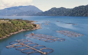
In BC, salmon production sites are defined as high or low risk based on a history of the occurrence, severity and outcomes of K. thyrsites infections ( Karreman et al., 2003).
Anecdotal information suggests that severity of infections among production salmon at high-risk sites can be lessened by prior residence of smolts at low-risk sites following transfer to the ocean (P. McKenzie, personal communication).
In this scenario, the transferred smolts will be larger upon first residence in high-risk sites therefore these observations do not distinguish between the possible effects of smolt size and prior exposure in contributing to the reduced risk of infection.
The purpose of this study was twofold: first to explore the possibility that prior exposure to the parasite increases the resistance of salmon to a subsequent exposure and second, to determine the effect of smolt size on the susceptibility of Atlantic salmon to K. thyrsites.
Discussion
Atlantic salmon were exposed to infective seawater by periodic cessation of UV-irradiation of RSW and first examined for infection with K. thyrsites 176 days or 2000 degree-days (DD) later.
In previous work, Atlantic salmon became infected with K. thyrsites following exposures to RSW from Departure Bay ( Moran et al., 1999b and Jones et al., 2012) and Moran et al. (1999c) showed that the peak plasmodium density occurred at approximately 25 weeks or 2000 DD. The efficacy of UV irradiation in negating the infectivity of seawater was most evident from the absence of infection in any of 40 UVSW controls in comparison to the 89% prevalence among 80 salmon that had been exposed to RSW and sampled at 176 day.
In addition, infections were not detected in 40 UVSW controls at 415 day. Previous research has shown that a UV dose of 44 mJ cm− 2 prevented Kudoa neurophila infections in Latris lineata ( Cobcroft and Battaglene, 2013).
Similarly, maximum UV doses of 68 and 216 mJ cm− 2 prevented Kudoa yasunagai and Kudoa amamiensis infections in Seriola lalandi and Seriola quinqueradiata, respectively ( Shirakashi et al., 2014). In a static freshwater system, a UV dose of 1300 mJ cm− 2 inactivated M. cerebralis triactinomyxons (TAMs) and prevented infections in juvenile rainbow trout at doses of 1400 and 14,000 TAMs fish− 1 (Hedrick et al., 2000).
In the present study, the rare detection of infection in UVSW controls sampled at 346 day may have been because of 10 system-level disruptions of UV-irradiation, each lasting from 10 min to 2 h. Despite this, our data indicate that a mean UV-irradiation of RSW at 350 mJ cm− 2 was effective at significantly reducing the number of K. thyrsites plasmodia in Atlantic salmon.
The present data showed that in Atlantic salmon, complete or partial recovery from K. thyrsites infection is associated with an increased resistance to subsequent homologous challenge. The prevalence and severity of K. thyrsites infections in Atlantic salmon declined between days 176 and 415, regardless of the duration of primary exposure.
Prevalence at the latter time was approximately 60% lower and median severity over 25-fold lower suggesting that in some fish, infections were resolved while in others they persisted at much reduced severity.
These observations agree with earlier researchers who found that K. thyrsites infection in laboratory-exposed Atlantic salmon are diminished 1year after initial exposure and that this recovery is associated with a chronic inflammation of the skeletal muscle ( Moran and Kent, 1999 and Moran et al., 1999c).
Following re-exposure at day 415, the prevalence and severity of infections among controls were elevated, similar to those observed following the initial exposure of naïve fish, whereas on average, infections among the previously exposed fish did not change from pre-exposure levels.
Recovery from infections with myxozoans or other parasites has been documented in salmonids and in the cases of Cryptobia salmositica, Ceratomyxa (= Ceratonova) shasta, L. salmonae, Gyrodactylus derjavini, Diplostomum spathaceum and Tetracapsuloides bryosalmonae, recovered fish are resistant to homologous challenge ( Jones and Woo, 1987, Bartholomew, 1998, Speare et al., 1998, Kent et al., 1999, Lindenstrøm and Buchmann, 2000, Karvonen et al., 2005 and Schmidt-Posthaus et al., 2012).
Similarly, infections with low-virulence strains of C. salmositica or L. salmonae increase host resistance against subsequent virulent homologous challenge, indicating vaccine potential ( Woo and Li, 1990 and Sanchez et al., 2001).
In the case of K. thyrsites, phagocytosis of myxospores and other parasite debris from ruptured plasmodia by macrophages during the recovery phase of infection ( Moran et al., 1999c) indicates immunological surveillance by the Atlantic salmon host. Similarly, increased severity of K. thyrsites during smoltification or sexual maturation in Atlantic salmon is inferred to be associated with depressed host immunity ( Moran and Kent, 1999 and St-Hilaire et al., 1998).
To date however, there are no data on mechanisms of the acquired protective immunity to K. thyrsites in recovered Atlantic salmon. Generally, defence mechanisms associated with the increased resistance to parasite challenge in recovered salmonids include transcription of innate and adaptive immune genes and production of non-specific and specific humoral and cellular factors ( Alvarez-Pellitero, 2008).
In assessing the effect of fish size on risk of infection, our results showed that fish exposed to RSW at 101 g had a significantly higher median severity of infection in comparison to heavier fish exposed at weights up to 332 g.
Contradicting this finding however, there was no statistically significant difference between the infection severity in the fish exposed at 101 g and controls in the first experiment exposed at 495 g (analysis not shown), suggesting size alone is not a useful predictor of risk.
Alternatively, we considered whether variable infection levels among the trials may have been related to seasonal differences in the infectivity of Departure Bay RSW, as suggested earlier by Moran and Kent (1999).
In that study, the parasite was detected in Atlantic salmon exposed in April, June, August or October but not in those exposed in December, January or March. Warmer water temperatures in Departure Bay in 2013–14 compared with 1995–96 (see Fig. 2 of Moran and Kent, 1999) may have been permissive to winter transmission, possibly explaining why we were able to detect infections throughout the year.
The abundance of K. thyrsites rDNA in RSW was measured using a quantitative PCR (qPCR) similar to that described earlier ( Funk et al., 2007). Evidence of significant variation among trials was observed, with the highest abundance of parasite rDNA occurring during trial 2. Coincidentally, the prevalence and severity of K. thyrsites infections were relatively low indicating that our RSW screening method was not a useful predictor of risk.
Despite this, seasonal analysis showed the abundance of parasite rDNA in Departure Bay was greatest among samples collected in summer and least in winter (not shown), in agreement with the previously observed pattern of RSW infectivity.
Poor agreement between the qPCR data and infectivity of RSW was likely related to inadequacies in our methods. For example, we screened at most 0.05% of the approximately 2.12 × 10⁶ L RSW flowing through each tank during intervals when UV-irradiation was discontinued, rendering the samples unlikely to be fully representative of the actual exposure. Furthermore, the qPCR is unable to distinguish between infective and non-infective parasite stages and likely therefore, to overestimate the risk of infection.
The identity of the K. thyrsites infective stage is not known and there is no information on relative abundances of putative actinospores and myxospores, the latter of which are not infective to naïve Atlantic salmon ( Moran et al., 1999b) but released into seawater during the decay of naturally-infected hosts.
Until we have a better understanding of actinospore structure and the ratio of actinospores to myxospores in RSW, there is little value in estimating actinospore concentrations from qPCR data.
Finally, although we demonstrated that the qPCR was reasonably specific, our samples may have included rDNA from other enzootic Kudoa spp. (i.e., K. paniformis) since a similar test amplified K. miniauriculata rDNA ( Funk et al., 2007). We will assess in ongoing work the extent to which rDNA abundance, obtained from a more frequent RSW sampling regime than used here, correlates with infection levels in concurrently exposed salmon.
In contrast to K. thyrsites in Departure Bay, the highest levels of K. yasunagai rDNA in Tanabe Bay, Japan occurred mid-winter, coincident with the coolest water temperatures ( Ishimaru et al., 2014).
Alternatively, peak concentrations of rDNA from Ceratomyxa puntazzi in the Mediterranean Sea, occurred in late summer to early autumn, somewhat later than the warmest water temperatures ( Alama-Bermejo et al., 2013). Similar approaches have been applied for the detection of DNA belonging to myxozoan fish pathogens in freshwater ( Hallett and Bartholomew, 2006 and Griffin et al., 2009).
In conclusion, the significant reduction in the prevalence and severity of K. thyrsites following re-exposure of previously infected salmon compared with controls, suggests opportunities for novel management strategies for soft-flesh in harvest-sized salmon. Such strategies may include the controlled early exposure of salmon to K. thyrsites as a form of vaccination. Further research is required to optimize this vaccine effect by studying the timing and duration of the primary exposure. In addition and with some refinement, the water-screening method described here will assist in the definition of risk of infection among production sites.