Introduction
Continuing the theme of diseases in tilapia we have highlighted in the previous two issues (Intervet Aquatic Animal Health Newsletter no. 11 and 12), another major bacterial disease known to have a devastating effect on survival rate in tilapia farms throughout the world is discussed here. The disease is highly contagious, especially at the fry and fingerling stages. Infected fish often display external lesions such as skin and gill erosion, and necrosis. In acute cases, these lesions may spread quickly and lead to high mortality within a matter of hours.
 Flavobacterium columnare on agar plate Flavobacterium columnare on agar plate |
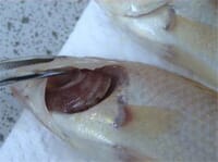
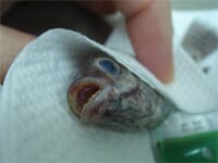 Mouth necrosis Mouth necrosis |
 Tail rot caused by columnaris Tail rot caused by columnaris |
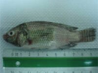 Saddleback lesion Saddleback lesion |
 Gill necrosis Gill necrosis |
F. columnare is a Gram negative, rod-shaped bacterium forming typical “hay stacks” or “columns” in wet-mount preparations (hence the name). These bacteria have a characteristic rhizoid pattern of growth on a low nutrient agar medium. Outbreaks are known to occur as a result of both temperature and environmental stress.
Clinical signs
External
Most columnaris infections are external manifested and present first as brown to yellowish-brown lesions on the gills, skin and fins. The lesions may first be seen only as a paler area that lacks the normal shiny appearance. These sores are usually surrounded by a zone with a distinct reddish tinge. Early signs of infection also include fin erosion.
Lesions on the back often extend down the sides, giving the appearance of a ‘saddle’, typical of columnaris disease. On the mouth, the lesions may look moldy or cottony, and the mouth can be severely affected. The gill lesions are typically necrotic and the filaments disintegrate as the bacteria invade them. Damage to the gills causes the fish to begin breathing rapidly and ‘gasping’ at the surface due to lack of oxygen.
Internal
Less commonly, the infection will be observed internally. During acute outbreaks, bacteria sometimes reach the blood system, resulting in a systemic infection.
Epidemiology
Fish are susceptible to columnaris following some degree of stress. Abrupt variations in water temperature are likely to induce and accelerate the progression of this disease. Poor water quality, inadequate diet, handling and overcrowding are also stress factors likely to induce an outbreak. Columnaris occurs frequently in fry production units (hatcheries) but also in cages and closed recirculation systems (grow-out facilities).
Once established, the disease is highly contagious and may spread horizontally from fish to fish, causing high mortality rates. Infections may also stem from the environment, through contaminated nets, specimen containers and even food. The presence of columnaris may also lead to secondary infection or other diseases; for example, winter saprolegniosis is often preceded by columnaris.
Diagnostic methods
A presumptive diagnosis is obtained by observation of typical clinical signs such as saddleback lesions, or necrotic gills and mouth. Additionally, the bacteria can be observed in a wet mount of infected tissues observed under light microscopy. Using phase contrast at 400x magnification, the bacteria show a slow gliding movement, gathering into characteristic column-like masses or “haystacks”.
A definitive diagnosis requires bacterial isolation on a low nutrient medium (such as cytophaga agar) and identification in the laboratory.
Control and treatment
The ideal way to eliminate the occurrence of columnaris is to alleviate stress in the cultured fish population. The bacteria thrive on organic wastes and these can be controlled by regular water changes. Proper diet, maintaining good water quality and avoidance of excessive handling will keep the fish from being stressed. To avoid spreading the bacteria, it is important to disinfect all equipment after each use and to use separate equipment at each rearing facility. But, this can be difficult in practice; however, stress should be minimized as much as possible. Salt (5-10 ppt) can be used to control the disease in hatchery tanks and to reduce the chance of infection during transportation.
In many cases, farmers are only able to partially control outbreaks by using antibiotics. However, this is not a sustainable practice. Best results are obtained if affected fish are treated as soon as the disease is detected. Infected fish display a reduced appetite and, consequently, antibiotics applied orally are generally ineffective. This may lead to selection of resistant bacteria leading to enhanced problems in the future. In general, antibiotics only prevent the infection from developing further. Farmers’ reports indicate that, once the infection is over, subsequent infections are less likely, indicating that an immune response can be elicited after infection.
A live-attenuated immersion vaccine, Aquavac-COLTM, against columnaris in channel catfish and manufactured by Intervet Inc. is on the market in the USA. In the future, we hope that a vaccine against F. columnare in tilapia and oher species will also be available as the key preventative technique to combat this devastating disease.
Further Information
Diseases of Tilapia - An Introduction
Streptococcus In Tilapia
February 2007