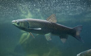
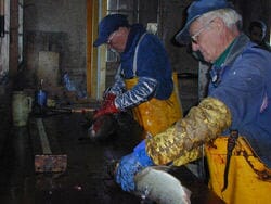
 Commercial fishers and biologists are concerned about the impact a viral disease will have on the Great Lakes fishery. There have been some large fish kills. Live fish commerce has been restricted to help prevent the spread of the disease. Commercial fishers and biologists are concerned about the impact a viral disease will have on the Great Lakes fishery. There have been some large fish kills. Live fish commerce has been restricted to help prevent the spread of the disease. |
Introduction
Viral hemorrhagic septicemia (VHS), a systemic infection of various salmonid and a few nonsalmonid fishes, is caused by a rhabdovirus designated as the viral hemorrhagic septicemia virus (Office International des Epizooties 1963). The virus infection occurs in salmonids and certain other fishes of any age and may result in significant cumulative mortality. Fish that survive may become carriers. Viral hemorrhagic septicemia has been reported under various names, of which "Egtved disease" and "Infektiose Nierenschwellung und Leberdegeneration" are the best known. The viral etiology of the disease was established by Jensen (1965), and several serotypes of the virus (also known as "Egtved virus") are currently recognized.
Comprehensive reviews of the disease and the virus have been published by de Kinkelin (1983), McAllister (1979), Pilcher and Fryer (1980), Roberts (1978), and Wolf (1988).
Geographical Distribution
Viral hemorrhagic septicemia is enzootic in most countries of continental Eastern and Western Europe, and the virus has been isolated in the Puget Sound area of Washington in the United States. No outbreaks of VHS or isolations of VHS virus have been reported elsewhere.
Host Susceptibility
In Europe, epizootics of VHS occur primarily in rainbow trout, Oncorhynchus mykiss; brown trout, Salmo trutta; and to a lesser extent in northern pike, Esox lucius (Jorgensen 1980; Meier and Jorgensen 1980). Natural infections have also occurred in grayling, Thymallus thymallus, and whitefish Coregonus sp. (Wizigmann et al. 1980; Ahne and Thomsen 1985; Meier et al. 1986). Outbreaks of VHS have been suspected in pollan, Coregonus lavaretus, and lake trout, Salvelinus namaycush. In the United States, natural infections have been diagnosed in chinook salmon, O. tshawytscha; coho salmon, O. kisutch; and steelhead (searun rainbow trout).
Fish shown by experimental challenge to be susceptible to VHS virus infection are Atlantic salmon, Salmo salar; brook trout, Salvelinus fontinalis; golden trout, O. aguabonita; rainbow trout x coho salmon hybrids; giebel, Carassius auratus gibelio; sea bass, Dicentrarchus labrax; and turbot, Scophthalmus maximus (de Kinkelin and Castric 1982; Castric and de Kinkelin 1984; Wolf 1988). Fish shown by experimental challenges to be refractory to VHS virus infection are common carp, Cyprinus carpio; chub, Leuciscus cephalus; Eurasian perch, Perca fluviatilis; roach, L. rutilus; and tench, Tinca tinca.
Clinical and Histopathologic Signs of Disease
A variety of clinical signs and histopathologic changes may be apparent in fish infected with VHS virus. Some fish show frank clinical manifestations of disease, whereas others look normal. Historically, clinical and pathologic signs of VHS have been catalogued into acute, chronic, and latent forms. Such descriptions represent degrees of severity rather than progressive stages of the disease.
Clinical Signs
The clinical signs of VHS vary with the severity of infection (Yasutake 1970; Wolf 1988). Acute signs are typically accompanied by a rapid onset of heavy mortality. Fish are lethargic, dark in color, exophthalmic, and anemic. Hemorrhages are evident in the eyes, skin, and gills and at the bases of the fins. Internally, punctiform hemorrhages are evident in periocular tissues, skeletal muscle, and viscera; the liver appears mottled and hyperemic and the kidneys are red and thin. In chronically infected fish, significant cumulative mortality occurs, but is protracted. Fish are lethargic, dark in color, exophthalmic, and severely anemic, but not grossly hemorrhagic. The abdomen is markedly distended due to edema of the liver, kidneys, and spleen. The liver appears pale and petechiated, and the kidneys are ashen. In a latent infection, mortality is low, and the fish seem nearly normal, but may be hyperactive. Inapparent virus carriers show no clinical signs of VHS.
Histopathological Changes
Histopathologic changes are generally confined to the liver, kidneys, spleen, and skeletal muscle (Ghittino 1965; Yasutake 1970; Amlacher et al. 1980; Wolf 1988). In acutely affected fish, the liver sinusoids are engorged with blood, and hepatocytes show focal to extensive necrobiotic changes-cytoplasmic vacuoles, pyknosis, karyolysis, Iymphocytic invasion, and occasionally intracytoplasmic and intranuclear inclusions. Similar changes occur in the spleen and in the hematopoietic and renal elements of the kidneys. In skeletal muscle, erythrocytes sometimes accumulate in muscle bundles and fibers, but little tissue damage occurs. In chronically infected fish, liver sinusoids remain enlarged and contain coagulated plasma, and kidney and splenic hematopoietic tissues and mononuclear lymphoid cells are hyperplastic. No remarkable histopathologic changes have been reported in inapparent virus carriers.
Etiology
The VHS rhabdovirion is an enveloped, bulletshaped particle about 180 nm long and 60 nm in diameter. Arrayed over the surface of the virion envelope are peplomers 5 to 15 nm long. The envelope is acquired as the virus matures, by budding from cell surface membranes or into cytoplasmic vacuoles. The intact virion, which sediments to a density of 1.141.15 g/cm3, is composed of five structural proteins and contains one segment of singlestranded RNA (de Kinkelin and Scherrer 1970; McAllister 1979; de Kinkelin 1983).
The virus is moderately stable in cell culture medium and can be preserved for years by freezing at 20°C or lower and by lyophilization. It is inactivated by exposure to ether, chloroform, glycerol, formalin, sodium hypochlorite, iodophors, ultraviolet irradiation, or heat (5660°C). The virus is stable at pH 5.010.4, but is inactivated at pH 3.5 (McAllister 1979; de Kinkelin 1983).
In cell culture, the VHS virus replicates to a titer of 107108 plaqueforming units per milliliter. The optimum temperature for virus replication is 1415°C; virus yield is reduced at 6°C, and little replication occurs above 20°C (de Kinkelin and Scherrer 1970). Optimum virus replication in cell culture requires that the pH of the medium be maintained in the range of pH 7.47.8 (Campbell and Wolf 1969). Interferon can be produced in cell cultures infected by VHS virus, and nascent or exogenous interferon reduces virus yield (de Kinkelin and Le Berre 1977).
At least three VHS virus serotypes (designated F1, F2, 23.75) can be distinguished by infectivity neutralization assays (Le Berre et al. 1977; de Kinkelin 1983). Hyperimmune sera for virus identification are generally prepared in rabbits, but virusneutralizing antibody titers are generally low because VHS virus is a weak immunogen (Ahne 1981; Hill et al. 1981). Hybridoma cell lines have been developed that secrete binding and neutralizing monoclonal antibody specific to VHS virus (Lorenzen et al. 1988; P. de Kinkelin, unpublished data; P. E. McAllister, unpublished data).
Virus Detection and Identification
The VHS virus can be recovered from homogenates of internal organs, sex products, or urine. Concentrations of virus are higher in the anterior kidney and spleen than in liver, heart, or muscle (de Kinkelin 1983). Brain samples should be included when one is assaying for inapparent virus carriers (Castric and de Kinkelin 1980). Little virus can be recovered from feces. Although infected fish can develop an immune response to VHS virus, the detection of antiVHS antibody is not a reliable indicator of the presence or absence of current infection (Dorson and Torchy 1979; Jorgensen 1982a,b). Nevertheless, the detection of VHS virusspecific neutralizing antibody in fish can be a useful tool for VHS surveillance (Olesen and Vestergard Jorgensen 1986). Mixed virus infections have been reported in which VHS virus was isolated from fish concurrently infected with infectious pancreatic necrosis virus.
The isolation of VHS virus in cell culture is the standard for diagnosis. The virus replicates in a variety of piscine cell lines-for example, BF2, CaPi, CHSE214, EPC, FHM, PG, RTG2, RTM, STE, and sea bass-as well as in primary cultures of tench, carp ovary, and goldfish, Carassius auratus (McAllister 1979; de Kinkelin 1983). In addition, VHS virus replicates in cell lines of mammalian (BHK21 and WI38) and reptilian (GL1 and TH1) origin. The optimum temperature for virus replication in cell culture is 1415°C (de Kinkelin and Scherrer 1970). Replication is greatly influenced by hydrogen ion concentration; a pH of 7.4 to 7.8 must be maintained in diagnostic assays (Campbell and Wolf 1969; Wolf and Quimby 1973). The detection of VHS virus in cell culture can be enhanced by pretreating cells with a 7% solution ofpolyethylene glycol (molecular weight = 20,000) or by including DEAEdextran (50mg/mL) or polyethylene glycol with the sample during virus adsorption (Campbell and Wolf 1969; W. N. Batts and J. R. Winton, unpublished data). Detection of virus in tissue sections by fluorescent antibody staining is less sensitive than virus isolation in cell culture (Jorgensen and Meyling 1972).
A variety of serological techniques can be used to identify VHS virus: infectivity neutralization, fluorescent antibody, immunoperoxidase, complement fixation, immune precipitation, countercurrent immunoelectophoresis, and immunoblot and plate enzymelinked immunosorbent assays (Meier and Vestergard Jorgensen 1975; Ahne 1981; de Kinkelin 1983; McAllister and Schill 1986; Way and Dixon 1988). The three currently recognized serotypes of VHS virus (F1, F2, and 23.75) are distinguished by infectivity neutralization; therefore, three antisera (or a composite polyvalent antiserum) are used for virus identification by infectivity neutralization assay. Viral antigen can be detected in inoculated cell cultures by indirect fluorescent antibody and immunoperoxidase staining (Meier and Jorgensen 1975; Faisal and Ahne 1980), and immunoblot and plate ELISA systems are used for detection and identification of VHS virus in cell culture fluids (McAllister and Schill 1986; Way and Dixon 1988).
Cross-reactivity is seen among the VHS virus serotypes in indirect fluorescent antibody assays and in some instances in infectivity neutralization assays (Jorgensen 1972, 1980; Meier and Jorgensen 1980). Similarly, one can identify all three VHS virus serotypes by immunoblot ELISA, using antiserum solely to VHS serotype F1 (McAllister and Owens 1987).
Epizootiology
Transmission
The VHS virus is readily transmissible to fish of all ages, and survivors of infection can become lifelong carriers that shed virus with urine and sex products. The virus ostensibly gains access to the fish through the secondary gill lamellae. Virus shed with sex products appears to be solely a surface contaminant of the egg and is readily dissipated. Although virus can be isolated from eggs for 3 to 4h after spawning, true vertical transmission has not been demonstrated. Experimentally, fish can be infected by cohabitation, immersion, intraperitoneal and intramuscular injection, brushing virus on the gills, and feeding (Jorgensen 1980; de Kinkelin and Castric 1982; de Kinkelin 1983; Castric and de Kinkelin 1984).
In Europe, the gray heron, Ardea cinerea, is known to be a mechanical vector of VHS virus (Olesen and Jorgensen 1982; Peters and Neukirch 1986), but the virus is inactivated in the gastrointestional tract of birds (Eskildsen and Jorgensen 1973). The virus is apparently not transmitted by parasitic vectors and, as judged from a study with the fruit fly Drosophila melanogaster, it does not replicate in insects (Bussereau et al. 1975).
In the hatchery environment, mechanical transfer of VHS virus on the surface of animate or inanimate objects presents a substantial hazard. The virus has been isolated from feral fish in waters receiving hatchery effluent, and can persist in water for several days (de Kinkelin and Scherrer 1970).
Factors Affecting the Disease
Fish of any age are susceptible to infection, although sac fry and fish older than 6 months are sometimes resistant, (Ghittino 1965). Epizootic losses occur at temperatures of 3° to 12°C (mortality is greatest at 3° to 5°C); mortality and the proportion of virus carriers decrease at higher temperatures (de Kinkelin 1983). Deaths from VHS rarely occur at temperatures above 15°C.
Infected fish mount a strong interferon response, but may ultimately succumb to infection (Dorson et al. 1975; de Kinkelin 1983). Increased interferon synthesis may play a role in transiently mitigating the effects of VHS at relatively high temperatures (de Kinkelin et al. 1982). Virusneutralizing antibody has also been demonstrated in infected fish, but the antibody response in survivors of epizootic and inapparent infections varies with the fish and the season of the year (Jorgensen 1971; Dorson and Torchy 1979; Dorson et al. 1979).
In captive fish, culture and environmental stress seemingly increase susceptibility and recurrence of infection (Ghittino 1965; de Kinkelin 1983). In feral fish, inapparent infection is more common than disease (Jorgensen 1982b) .
Disease Control
Prevention of contact between the virus and the host is the most effective method for controlling VHS. A systematic program of hatchery disinfection, combined with restocking with specificpathogenfree fish and eggs, has been used successfully (Kehlet 1973; Jorgensen 1974a, 1980). Eggs used for restocking are decontaminated by iodophor treatment. The water supply should ideally be controlled and virusfree, although ultraviolet irradiation has been used to inactivate VHS virus in the water supply (Maisse et al. 1980). Conditions that promote physiological stress should be alleviated. Although VHS rarely occurs above 15°C, disease control by temperature manipulation has not been described. Selective breeding to increase host resistance to VHS has not been successful.
The potential for vaccinating fish against VHS has been demonstrated, and several avirulent VHS virus vaccines are under development (Jorgensen 1976; de Kinkelin and Le Berre 1977). One vaccine candidate, a VHS virus variant of low pathogenicity (F25), was selected by serial passage in EPC cells at progressively increasing temperature. This F25 variant replicates at 25°C, but the wildtype virus does not (de Kinkelin et al. 1980; de Kinkelin and Bearzotti 1981). A second vaccine candidate, a low-pathogenicity virus strain (Reva), was selected by serial passage in RTG2 cells (Jorgensen 1982b). Although both the F25 and the Reva strains induced a protective response, they also retained residual pathogenicity. The mechanism of the protective response is unclear. Some mechanism occurring within 48h after immunization, such as interferon stimulation, might be responsible for early protection, whereas the antibody response occurs later (Jorgensen 1982b; Bernard et al. 1985). Fry can be protected by injection of interferon (de Kinkelin et al. 1982). Although the protective effects of avirulent vaccines have been demonstrated in the laboratory, their efficacy under production conditions has not been proven, and their value for controlling VHS in healthy fish populations has been questioned (Erlzmann 1983).
Bibliography
Ahne, W. 1981. Serological techniques currently used in fish virology. Dev. Biol. Stand. 49:327.Ahne, W., and I. Thomsen. 1985. Occurrence of viral hemorrhagic septicemia virus in wild whitefish Coregonus sp. Zentralbl. Veterinaermed. Reihe B 32:7375.
Amlacher, E., J. Ude, C. Rudolph, and G. Ernst. 1980. Direct electron microscopial visualization of the presumptive virus of viral haemorrhagic septicaemia (VHS) in rainbow trout Salmo gairdneri Richardson and additional histopathological and haematological observations. J. Fish Dis. 3:5562.
Bernard, J., M. BearzottiLe Berre, and P. de Kinkelin. 1985. Viral haemorrhagic septicaemia in rainbow trout: attempt to relate interferon production, antibody synthesis and structure of the virus with the mechanism of virulence. Ann. Inst. Pasteur/Virol. 136: 1326.
Campbell, J. B., and K. Wolf. 1969. Plaque assay and some characteristics of Egtved virus (virus of viral hemorrhagic septicemia of rainbow trout). Can. J. Microbiol. 15:635637.
Castric, J., and P. de Kinkelin. 1980. Occurrence of viral haemorrhagic septicaemia in rainbow trout Salmo gairdneri Richardson reared in seawater. J. Fish Dis. 3:2127.
Castric, J., and P. de Kinkelin. 1984. Experimental study of the susceptibility of two marine fish species, sea bass (Dicentrarchus labrax) and turbot (Scophthalmus maximus), to viral haemorrhagic septicaemia. Aquaculture 41:203212.
de Kinkelin, P. 1983. Viral haemorrhagic septicaemia. Pages 5162 in D. P. Anderson, M. Dorson, and Ph. Dubourget, eds. Antigens of fish pathogens. Fondation Marcel Merieux, Lyon, France.
de Kinkelin, P., and M. Bearzotti. 1981. Immunization of rainbow trout against viral hemorrhagic septicemia (VHS) with a thermoresistant variant of the virus. Proceedings of the International Symposium on Fish Biologics: Serodiagnostics and Vaccines, National Fish Health Research Laboratory, Leetown, W. Va., 2630 April 1981.
de Kinkelin, P., M. BearzottiLe Berre, and J. Bernard. 1980. Viral hemorrhagic septicemia of rainbow trout: selection of a thermoresistant virus variant and comparison of polypeptide synthesis with the wildtype virus strain. J. Virol. 36:652658.
de Kinkelin, P., and J. Castric. 1982. An experimental study of the susceptibility of Atlantic salmon fry, Salmo salar L., to viral haemorrhagic septicaemia. J. Fish Dis. 5:5765.
de Kinkelin, P., M. Dorson, and A. M. HattenbergerBaudouy. 1982. Interferon synthesis in trout and carp after viral infection. Pages 167174 in W. B. van Muiswinkel and E. L. Cooper, eds. Immunology and immunization of fish. Pergamon Press, New York.
de Kinkelin, P., and M. Le Berre. 1977. Isolement d'un Rhabdovirus pathogene de la Truite Fario (Salmo trutta, L. 1766). C. R. Acad. Sci. Paris 284D:101104.
de Kinkelin, P., and R. Scherrer. 1970. Le Virus d'Egtved I. Stabilite, developpement et structure du virus de la souche danoise F1. Ann. Rech. Vet. 1:1730.
Dorson, M., A. Barde, and P. de Kinkelin. 1975. Egtved virus induced rainbow trout serum interferon: some physiochemical properties. Ann. Microbiol. 126B:485489.
Dorson, M., and C. Torchy. 1979. Complement dependent neutralization of Egtved virus by trout antibodies. J. Fish Dis. 2:345347.
Dorson, M., C. Torchy, and C. Michel. 1979. Rainbow trout (Salmo gairdneri) complement fixation used for titration of antibodies against several pathogens. Ann. Rech. Vet. 10:529534.
Enzmann, P. J. 1983. Considerations on the effectiveness of VHSvaccination. Bull. Eur. Assoc. Fish Pathol. 5:5455.
Ghittino, P. 1965. Viral hemorrhagic septicemia (VHS) in rainbow trout in Italy. Ann. N.Y. Acad. Sci. 126:468478.
Hill, B. J., R. F. Williams, and J. Finlay. 1981. Preparation of antisera against fish virus disease agents. Dev. Biol. Stand. 49:209218.
Jensen, M. H. 1965. Research on the virus of Egtved disease. Ann. N.Y. Acad. Sci. 126:422426.
Jorgensen, P. E. V. 1974a. A study of viral diseases in Danish rainbow trout, their diagnosis and control. Commissioned by A/S Carl Fr. Mortensen, Bulowsvej 5 C 1870 Kobenhavn V, Denmark, 101 pp.
Jorgensen, P. E. V. 1974b. Indirect fluorescent antibody techniques for demonstration of trout viruses and corresponding antibody. Acta Vet. Scand. 15:198205.
Jorgensen, P. E. V. 1976. Partial resistance of rainbow trout (Salmo gairdneri) to viral haemorrhagic septicaemia (VHS) following exposure to nonvirulent Egtved virus. Nord. Veterinaermed. 28:570571.
Jorgensen, P. E. V. 1980. Egtved virus: the susceptibility of brown trout and rainbow trout to eight virus isolates and the significance of the findings for the VHS control. Pages 37 in W. Ahne, ed. Fish diseases. Third COPRAQSession. SpringerVerlag, Berlin, Heidelberg, New York.
Jorgensen, P. E. V. 1982a. Egtved virus: occurrence of inapparent infections with virulent virus in freeliving rainbow trout, Salmo gairdneri Richardson, at low temperature. J. Fish Dis. 5:251255.
Jorgensen, P. E. V. 1982b. Egtved virus: temperaturedependent immune response of trout to infection with lowvirulence virus. J. Fish Dis. 5:4755.
Jorgensen, P. E. V., and A. Meyling. 1972. Egtved virus: demonstration of virus antigen by the fluorescent antibody technique in tissues of rainbow trout affected by viral haemorrhagic septicaemia and in cell cultures infected with Egtved virus. Arch. Gesamte Virusforsch. 36:115122.
Kehlet, N. P. 1973. A summary of the rules, methods and results of the Danish campaign against infectious disease of freshwater fish. EIFAC [Eur. Inland Fish. Advis. Comm.] Tech. Pap. 17, Suppl. 2:3738.
Le Berre, M., P. de Kinkelin, and A. Metzger. 1977. Identification serologique des rhabdovirus des salmonides. Bull Off. Int. Epizoot. 87:391393.
Lorenzen, N., N. J. Olesen, and P. E. Vestergard Jorgensen. 1988. Production and characterization of monoclonal antibodies to four Egtved virus structural proteins. Dis. Aquat. Org. 4:3542.
Maisse, G., M. Dorson, and C. Torchy. 1980. Ultraviolet inactivation of two pathogenic salmonid viruses (IPN virus and VHS virus). Bull. Fr. Piscic. 278:3440.
McAllister, P. E. 1979. Fish viruses and viral infections. Pages 401470 in H. FraenkelConrat and R. R. Wagner, eds. Comprehensive virology, Vol. 14. Plenum Publishing Corp., New York.
McAllister, P. E., and W. J. Owens. 1987. Identification of the three serotypes of viral hemorrhagic septicemia virus by immunoblot assay using antiserum to serotype Fl. Bull. Eur. Assoc. Fish. Pathol. 7:9092.
McAllister, P. E., and W. B. Schill. 1986. Immunoblot assay: a rapid and sensitive method for identification of salmonid fish viruses. J. Wildl. Dis. 22:468474.
Meier, W., and P. E. V. Jorgensen. 1975. A rapid and specific method for the diagnosis of viral haemorrhagic septicaemia (VHS) of rainbow trout. Riv. Ital. Piscic. Ittiop. 10:1115.
Meier, W., and P. E. V. Jorgensen. 1980. Isolation of VHS virus from pike fry (Esox lucius) with hemorrhagic symptoms. Pages 817 in W. Ahne, ed. Fish diseases. Third COPRAQSession. SpringerVerlag, Berlin, Heidelberg, New York.
Meier, W., W. Ahne, and P. E. V. Jorgensen. 1986. Fish viruses: Viral haemorrhagic septicaemia in whitefish (Coregonus sp.). J. Appl. Ichthyol. 4:181186.
Office International des Epizooties. 1963. Resolutions. Bull. Off. Int. Epizoot. 59:291295.
Olesen, N. J., and P. E. V. Jorgensen. 1982. Can and do herons serve as vectors for Egtved virus? Bull. Eur. Assoc. Fish Pathol. 2:48.
Olesen, N. J., and P. E. Vestergard Jorgensen. 1986. Detection of neutralizing antibody to Egtved virus in rainbow trout (Salmo gairdneri) by plaque neutralization test with complement addition. J. Appl. Ichthyol. 2:3341.
Peters, F., and M. Neukirch. 1986. Transmission of some fish pathogenic viruses by the heron, Ardea cinerea. J. Fish Dis. 9:539544.
Pilcher, K. S., and J. L. Fryer. 1980. The viral diseases of fish: a review through 1978. Part 1: Diseases of proven viral etiology. Rev. Microbiol. 7:287364.
Roberts, R. J., editor. 1978. Fish pathology. Bailliere Tindall, London. 319 pp.
Vestergard Jorgensen, P. E. 1972. Egtved virus: antigenic variation in 76 virus isolates examined in neutralization tests and by means of the fluorescent antibody technique. Symp. Zool. Soc. Lond. 30:333340.
Way, K., and P. E. Dixon. 1988. Rapid detection of VHS and IHN viruses by the enzymelinked immunosorbent assay (ELISA). J. Appl. Ichthyol. 4:182189.
Wizigmann, G., C. Baath, and R. Hoffmann. 1980. Isolation of viral hemorrhagic septicemia virus from fry of rainbow trout, pike, and grayling. Zentralbl. Veterinaermed. Reihe B 27:7981.
Wolf, K. 1988. Fish viruses and fish viral diseases. Cornell University Press, Ithaca, N.Y. 476 pp.
Wolf, K., and M. C. Quimby. 1973. Fish viruses: buffers and methods for plaquing eight agents under normal atmosphere. Appl. Microbiol. 25:659664.
Yasutake, W. T. 1970. Comparative histopathology of epizootic salmonid virus diseases. Pages 341350 in S. F. Snieszko, ed. A symposium on diseases of fishes and shellfishes. Am. Fish. Soc. Spec. Publ. 5.
By Philip E. McAllister - U.S. Fish and Wildlife Service, National Fisheries Research Center-Leetown, National Fish Health Research Laboratory, Box 700, Kearneysville, West Virginia. 25430.
Further Information
For additional information on Viral Haemorrhagic Septicaemia by the OIE, click here
or read Cefas's Viral Haemorrhagic Septicaemia Fact Sheet by clicking here
Added January 2007