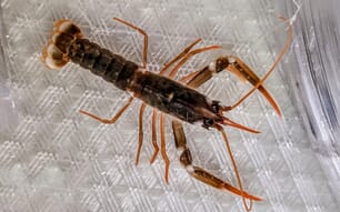
The giant freshwater prawn, Macrobrachium rosenbergii is a native inhabitant of sub-tropical and tropical waters. It is commercially important and one of the most cultured prawn species in Southeast Asia, as well as Israel, Japan, Taiwan Province of China, Latin America, the Caribbean and some countries in Africa due to its potential of fast growth, large size, disease tolerance and export market value [1].
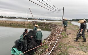
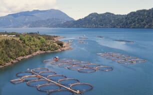
Increasing demand for this species in both the domestic and export markets has led to a remarkable increase in the number of large scale culture systems with high stocking density and intensive feeding [2]. With the rapid development in hatchery production of post-larvae and the number of prawn grow-out farms, good husbandry and environmental management have often been neglected. Consequently, pathogens gain easy entry as the prawns are stressed and weakened under adverse environmental conditions [3].
Diseases of viral aetiology have caused havoc in the aquaculture industry, sometimes wiping out entire stocks within days of onset of infection [4]. Macrobrachium rosenbergii is often considered as a less susceptible species to disease problems when compared with farmed penaeid shrimp, perhaps because of the generally less intensified culture practices of freshwater prawn farming. However, recently disease problems have become of serious concern to giant freshwater prawn farming, probably because of the intensification of culture and the translocation of seed and broodstock [5]. The recent report of white muscle disease (WMD) in freshwater prawn hatcheries and farms has sent shock waves through the prawn aquaculture industry.
Whitish muscle disease (WMD)
Whitish muscle disease of Macrobrachium rosenbergii, also called "whitish disease" or "white tail disease", is a new serious epizootic disease caused by Macrobrachium rosenbergii nodavirus (MrNV). It was first observed and reported in a hatchery in Guadeloupe, French West Indies [6]. Then later on in China (both mainland and the Province of Taiwan) and India. It has caused immense economic losses in hatcheries and farms, with mortalities often reaching 100 per cent in post-larval stages [10,13]. These viruses thrive in brackish and freshwater environment. The specific host is giant freshwater prawn and the affected life stages are larvae, post-larvae and early juveniles [7]. There is no evidence of adult life stages being affected but they might act as carriers. It has not been listed by OIE.
The geographical distribution of this virus is across northern South America (French West Indies, Dominican Republic and Caribbean region) and Asia (China, Taiwan Province of China and India). Clinical signs and mortality patterns appear similar in all locations and it may be assumed that movement of some common prawn population source might be the reason for the wide distribution of the WMD. However, further studies are required to understand the geographic distribution [7].
Pathogen and mode of infection
The causative agents of WMD are thought to be M. rosenbergii nodavirus (MrNV) and extra small virus (XSV). These two RNA viruses are invariably seen in association with infected prawns. MrNV is a small icosahedral non-enveloped virus, 26 to 27 nm in diameter that has been identified in the cytoplasm of connective tissue cells. The capsid contains a single polypeptide of 43 kDa (cp-43) [8]. The nodaviruses are known to contain a genome consisting of two single-stranded positive-sense RNA segments, RNA1, which encodes the viral part of the RNA-dependent RNA polymerase (RdRp) and RNA2, which encodes the capsid protein. In infected cells, RNA3, a subgenomic transcript of RNA1, is also present [9]. Viral replication takes place in the cytoplasm of connective tissue cells of most organs and tissues. XSV is also an icosahedral virus, 15 nm in diameter, its genome consisting of a linear single-stranded positive-sense RNA coding for a capsid protein, cp-17. Because of its extremely small size and absence of gene-encoding enzymes required for replication, it has been suggested that XSV may be a satellite virus, while MrNV plays the role of a helper virus [10]. However, the respective roles of the two viruses in the disease pathogenesis are not yet known11. This disease outbreak occurs in post-larvae 3–5 days to 3 weeks after desalting. Incidence of post-larval mortalities of 30-100 per cent within two or three days of appearance of the clinical sign of opaqueness was reported [5]. Very few post-larvae presenting these signs survive and survivors seem to grow normally in grow-out ponds [7]. Bacteriological examination of affected PL showed the presence of Staphylococcus spp. as a predominant organism, while laboratory challenge of healthy PL with this bacterial isolate did not reproduce WMD [5].
Signs and symptoms
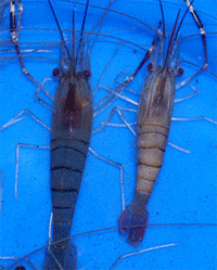
The clinical signs of this disease include lethargy, anorexia and opaqueness of abdominal muscle in post-larvae and adults. Whitish appearance of the tail is the prominent clinical sign, and therefore, the disease is also named as white tail disease. This milky opaqueness gradually expands on both sides (anterior and posterior) and leads to degeneration of telson and uropods in severe cases. Some infected animals without uropods have been observed. The discoloration appears to start at the tail extremity (telson region) and gradually progress towards the head. Eventually all muscles in the abdomen and cephalothorax are affected. The tissues most affected in moribund PLs/early juveniles are striated muscles of the abdomen and cephalothorax and intratubular connective tissue of the hepatopancreas [7]. The cephalothoracic region increases in size and it may become double the original size (referred to as branchiostegite blister disease (BBD) or swollen head syndrome). The cephalothoracic region when opened, may contain two sac-like structures with watery fluid above the hepatopancreas on either side. Histopathological examination of the infected animals reveals highly necrotic musculature. Multifocal areas of hyaline necrosis of muscle fibres are found in the striated muscle [7]. Degenerated muscle areas show aggregations of melanized nuclei, many of which look like inclusion bodies. The clinical signs and histopathology of WMD closely resemble to the idiopathic muscle necrosis (IMN) reported in M. rosenbergii [12].
Diagnostic methods
To curb the disease spread and to avoid economic loss, it is essential that a highly sensitive, specific and rapid diagnostic method be developed for early detection of both the pathogenic agents (MrNV and XSV) of the disease. The three basic methods in disease diagnosis are screening, presumptive and confirmatory methods [7]. The presumptive method includes gross observation of the presence of post-larvae with milky white colour abdomen followed by mortality, histopathological study of changes characterized by pale to darkly basophilic, reticulated cytoplasmic inclusions in the connective tissue cells of most organs and tissues (pryonin methyl green staining can be used to distinguish the characteristically green-stained MrNV viral inclusions from hemocyte nuclei) and virological studies. The screening and confirmatory tests can be done using viral genome-based detection methods using reverse-transcriptase polymerase chain reaction (RT-PCR) and loop-mediated isothermal amplification (LAMP). Other detection methods for MrNV include a double antibody sandwich enzyme-linked immunosorbent assay (DS-ELISA), triple antibody sandwich enzyme-linked immunosorbent assay (TAS-ELISA) and dot blot hybridization, in situ hybridization.
Reverse-transcriptase polymerase chain reaction
RT-PCR is the most sensitive of all diagnostic method to detect MrNV. This method is used to synthesis and amplifiy cDNA copies from RNA viruses. Identification of virus can be done using amplification of cDNA by specific primers. The primer sequence for MrNV is 5’-GCG-TTA-TAG-ATG-GCA-CAA-GG-3’ (forward) and 5’-AGC-TGT-GAA-ACT-TCC-ACT-GG-3’ (reverse) with amplified product size of 425 bp [7]. Although very sensitive and highly specific, it requires the use of a thermal cycler and hence can be carried out only in well-equipped laboratories. More recently a single-tube, duplex RT-PCR method has been developed for simultaneous detection of MrNV and XSV.
Loop-mediated isothermal amplification
A loop-mediated isothermal amplification (LAMP) procedure is described for rapid diagnosis of white muscle disease. It is a specific nucleic acid amplification method that can amplify target nucleic acid to 109 copies at 60–65°C in 1 hour [13]. The method relies on autocycling strand displacement DNA synthesis by the best DNA polymerase large fragment, a DNA polymerase with high strand displacement activity. As the reaction is carried out under isothermal conditions, it can be performed with a simple and inexpensive water bath. As there is no time loss in thermal changes, the amplification efficiency of the LAMP method is extremely high. The LAMP reaction requires four primers specifically recognizing six distinct regions of the template DNA. Hence, there is a high degree of specificity for detection. A positive reaction can be easily detected within one hour due to the production of a whitish precipitate of magnesium pyrophosphate thereby eliminating the need for agarose gel electrophoresis [14]. The time kinetics and sensitivity of the LAMP reaction can be further improved by the use of two additional loop primers. There are very few reports of the application of LAMP methodology for detection of RNA viruses. An RT-LAMP method has been developed as a rapid and convenient method for detection.
Transmission
Both vertical and horizontal transmission of MrNV has been observed [7]. Infected broodstock serve as carriers and results in diseased postlarvae. Even Artemia nauplii from infected Artemia stocks can spread the disease. Penaeid shrimps are not much susceptible to these viruses but experimental results indicate the possibility of shrimp acting as reservoir for MrNV and maintaining their virulence in their tissue system.
Conclusion
Prevention is better than cure, as there are no treatments to this viral pathogen, only through adoption of better management practices in hatcheries and farms can the spread and impact of white tail disease in prawn farming can be minimized. The early screening of broodstock and postlarvae should be strongly encouraged. Strict quarantine measures should is followed in translocation of prawn stock and seeds to avoid spread of pathogen. Broodstock or seed testing positive for MrNV must be discarded with proper zoosanitory methods. Further research is needed for through understanding of pathogen-host interaction, viral strain, immune response and drug development.
References
1. New, M.B. (1990). Freshwater prawn culture: a review. Aquaculture 88: 99-143.
2. Phuong, N.T., Tuan, N.A., Hien, T.T.T., Hai, T.N., Wilder, M., Ogata, H., Sano, M. and Maeno, Y. (2002). Development of freshwater prawn (Macrobrachium rosenbergii) seed production and culture technology in the Mekong delta region of Vietnam: A review of the JIRCAS project at Cantho University.
3. Tonguthai, K. (1997) Diseases of the Freshwater Prawn, Macrobrachium rosenbergii. AAHRI Newsletter 4.2.
4. Pillai, D., Bonami, J.R, and Sri Widada, J. (2006). Rapid detection of Macrobrachium rosenbergii nodavirus (MrNV) and extra small virus (XSV), the pathogenic agents of white tail disease of Macrobrachium rosenbergii (De Man), by loop-mediated isothermal amplification. J. Fish Diseases 29: 275–283.
5. Vijayan, K.K., Stalin Raj, V., Alavandi, S.V., Sekhar, V.T and Santiago, T.C. (2005). Incidence of white muscle disease, a viral like disease associated with mortalities in hatchery-reared postlarvae of the giant freshwater prawn Macrobrachium rosenbergii (De Man) from the southeast coast of India. Aquaculture Research 36: 311-316.
6. Arcier, J.M., Herman, F., Lightner, D.V., Redman, R.M., Mari, J. and Bonami, J.R. (1999). A viral disease associated with mortalities in hatchery-reared postlarvae of the giant freshwater prawn Macrobrachium rosenbergii. Dis. Aquat. Org. 38: 177-181.
7. Sahul Hameed, A.S. (2005). White tail disease card developed to support the NACA/FAO/OIE regional quarterly aquatic animal disease (QAAD) reporting system in the Asia-Pacific. NACA, Bangkok, Thailand. 7 pp.
8. Bonami, J.R., Shi, Z., Qian, D. and Sri Widada, J. (2005). White tail disease of the giant freshwater prawn, Macrobrachium rosenbergii: separation of the associated virions and characterization of MrNV as a new type of nodavirus. Journal of Fish Diseases 28: 23–31.
9. Sommerset, I. and Nerland, A.H. (2004). Complete sequence of RNA1 and subgenomic RNA3 of Atlantic halibut nodavirus (AHNV). Diseases of Aquatic Organisms 58: 117–125.
10. Sahul Hameed, A.S., Yoganandhan, K., Sri Widada, J. and Bonami, J.R. (2004). Studies on the occurrence and RT-PCR detection of Macrobrachium rosenbergii nodavirus (MrNV) and extra small virus like particles (XSV) associated with white tail disease (WTD) of Macrobrachium rosenbergii in India. Aquaculture 238: 127–133.
11. Sri Widada, J., Richard, V., Cambournac, I., Shi, Z., Qian, D. and Bonami, J.R. (2004) Dot-blot hybridisation and RT-PCR detection of extra small virus (XSV) associated with white tail disease of prawn Macrobrachium rosenbergii. Diseases of Aquatic Organisms 58: 83–87.
12. Nash, G., Chinabut, S. and Limsuwan, C. (1987). Idiopathic muscle necrosis in the freshwater prawn, Macrobrachium rosenbergii de Man, cultured in Thailand. Journal of Fish Diseases 10: 109-120.
13. Notomi, T., Okayama, H., Masubuchi, H., Yonekawa, T., Watanabe, K., Amino, N. and Hase, T. (2000). Loop-mediated isothermal amplification reaction of DNA. Nucleic Acids Research 28: 63.
13. Sri Widada, J., Durand, S., Cambournac, I., Qian, D., Shi, Z., Dejonghe, E., Richard, V. and Bonami, J.R. (2003). Genome based detection methods of Macrobrachium rosenbergii nodavirus, a pathogen of the giant freshwater prawn, Macrobrachium rosenbergii: dot-blot, in situ hybridization and RT-PCR. Journal of Fish Diseases 26: 583–590.
14. Mori, Y., Nagamine, K., Tomita, N. and Notomi, T. (2001). Detection of loop-mediated isothermal amplification reaction by turbidity derived from magnesium pyrophosphate formation. Biochemical and Biophysical Research Communications 289:, 150–154.
15. Sri Widada, J. and Bonami, J.R. (2004). Characteristics of the monocistronic genome of extra small virus, a virus-like particle associated with Macrobrachium rosenbergii nodavirus: possible candidate for a new species of satellite virus. Journal of General Virology 85: 643–646.
February 2009